Elbow reconstruction
Elbow surgery
Arthroscopic and open treatment for instability, tendon injury, stiffness, arthritis, trauma, and throwing-related problems.
The elbow combines a stable hinge with forearm rotation and is surrounded by important nerves, ligaments, and tendons. Surgery is individualized to the diagnosis and may be arthroscopic, open, or a combination of both.
See the anatomy and imaging
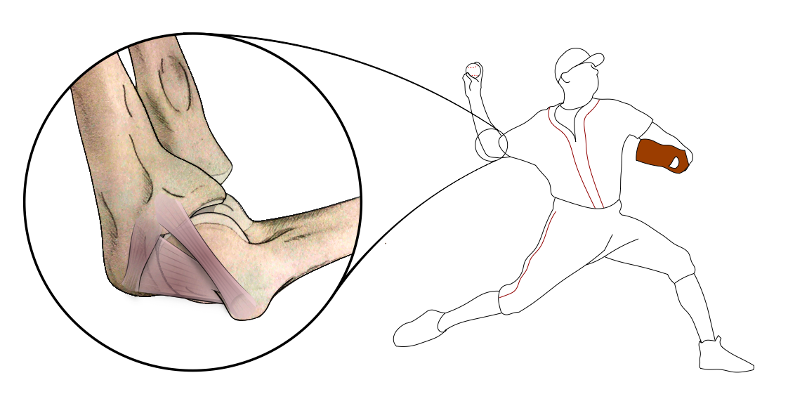
Anatomy that guides treatment
The ulnohumeral joint acts as a hinge, while the radiocapitellar and proximal radioulnar joints help the forearm rotate. The medial and lateral ligaments stabilize the elbow; the biceps and triceps power flexion and extension; and the ulnar, median, and radial nerves pass close to the surgical field.
Conditions treated
- Loose bodies, stiffness, and selected arthritis
- Tennis elbow and distal biceps or triceps injury
- Ulnar collateral ligament injury in throwers
- Fractures, instability, and post-traumatic problems
- Selected cases requiring elbow replacement
Arthroscopic procedures
Elbow arthroscopy can remove loose bodies, release scar tissue, treat selected cartilage or impingement problems, and assist evaluation of the joint. Because major nerves lie close to the portals, careful positioning, anatomy, and technique are especially important.
Open repair and reconstruction
Open surgery may repair the distal biceps or triceps, reconstruct a collateral ligament, stabilize a fracture, release severe stiffness, decompress or transpose a nerve, or replace a damaged joint. UCL repair with internal brace or reconstruction is selected according to tear location, tissue quality, athlete goals, and chronicity.
Imaging and planning
X-rays assess arthritis, alignment, fractures, and loose bodies. CT defines complex bone anatomy and joint-surface injury. MRI or ultrasound may be used for tendons, ligaments, nerves, and cartilage. Electrodiagnostic testing is sometimes useful when nerve compression or injury is suspected.
Recovery
The plan depends on whether bone, ligament, tendon, or cartilage was treated. Early controlled motion is often important, but repaired structures also need protection. Your splint, brace, lifting limits, and therapy schedule are procedure specific.
Risks and realistic goals
The elbow can become stiff and its nearby nerves can be irritated. Other risks include infection, instability, incomplete healing, heterotopic bone, persistent pain, weakness, hardware problems, and repeat surgery. The aim is pain relief, stability, useful motion, and safe return to work or sport; normal motion is not guaranteed after every injury.
Evidence and further reading
- AAOS OrthoInfo: Elbow Arthroscopy
- AAOS OrthoInfo: Ulnar Collateral Ligament Injury
- AAOS OrthoInfo: Distal Biceps Tendon Tear
Educational content is reviewed for patient clarity and is not a substitute for an examination or individualized medical advice.
PIA Shoulder MD